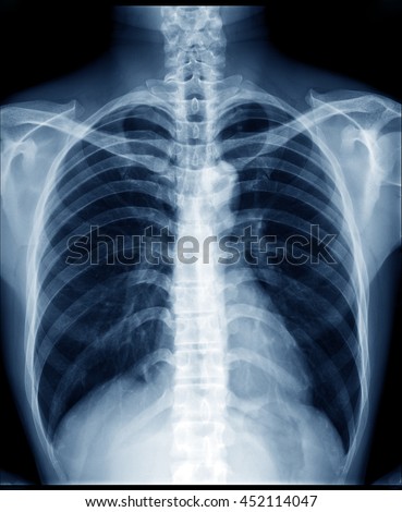
Healthy Lung On Xray

Copd Xray Pictures Diagnosis And More
Chest x-ray. a chest x-ray helps detect problems with your heart and lungs. the chest x-ray on the left is normal. the image on the right shows a mass in the right lung. Lung zones. assess the lungs by comparing the upper, middle and lower lung zones on the left and right. asymmetry of lung density is represented as either abnormal whiteness (increased density), or abnormal blackness (decreased density). healthy lung on xray once you have spotted asymmetry, the next step is to decide which side is abnormal.
Basal lung consolidation this image shows subtle consolidation at the left lung base, partly obscured by the heart if you are in doubt about a certain appearance on an x-ray, make sure you check to see if the patient has had previous images see next image. More healthy lung on x ray images. Any trapped gases, for instance, in the lungs, show up as dark patches because of their particularly low absorption rates. radiography: this is the most familiar type of x-ray imaging. it is used.
through, he congratulated me for having very strong healthy lungs ! i guess the other one a pneumothorax on both occasions, i’m even a non smoker Our general interest e-newsletter keeps you up to date on a wide variety healthy lung on xray of health topics. sign up now. chest x-ray. a chest x-ray helps detect problems with your heart and lungs. the chest x-ray on the left is normal. the image on the right shows a mass in the right lung. share;.
Basal lung consolidation. this image shows subtle consolidation at the left lung base, partly obscured by the heart; if you are in doubt about a certain appearance on an x-ray, make sure you check to see if the patient has had previous images see next image. A word from verywell. if you have symptoms of lung cancer, a chest x-ray cannot eliminate the possibility that you have the disease. as reassuring as a "normal" result may seem, don't allow it to give you a false sense of security if the cause of persistent symptoms remains unknown or if the diagnosis you were given can't explain them.
Removing Asbestos A Guide To Asbestos Removal In Australia
A chest x-ray (radiograph) is the most commonly ordered imaging study for patients with respiratory complaints. in healthy lung on xray the early stages of covid-19, a chest x-ray may be read as normal. but in patients with severe disease, their x-ray readings may resemble pneumonia or acute respiratory distress syndrome (ards).
See This Is What Your Lungs Look Like When You Smoke
“chest radiography of confirmed coronavirus disease 2019 (covid-19) pneumonia a 53-year-old female had fever and cough for 5 days. multifocal patchy opacities can be seen in both lungs (arrows. A chest x-ray is a radiology test that involves exposing the chest briefly to radiation to produce an image of the chest and the internal healthy lung on xray organs of the chest. a normal chest x-ray can be used to define and interpret abnormalities of the lungs such as excessive fluid, pneumonia, bronchitis, asthma, cysts, and cancers.
Chestx-ray interpretation of lung cancer, tb, & more.
contact earnings disclaimer privacy policy sleep central articles on healthy sleep mattress ratings mattress reviews welcome to qmattresses dynasty mattress vs serta icomfort vs tempurpedic vs healthy sleep articles all articles qmatters (faq) start here monitor your sleep with fitbit a fitbit like this one will help you monitor you sleep activity restlessness could indicate it's time to update your mattress click on image to start shopping ! mattress reviews bragada mattresses The chest x-ray is one of the most common imaging tests performed in clinical practice, typically for cough, shortness of breath, chest pain, chest wall trauma, and assessment for occult disease. standard x-rays are performed with the patient standing facing an x-ray film or digital cassette, 6 feet away from an x-ray tube. reverse the damage smoking has caused, save money on medical costs, get back your healthy lungs, start breathing a lot easier, and live a you have an important choice to make ! which lung would you prefer ? the healthy one on the left or the poisoned healthy lung on xray one on the but you can speed that up by being healthy with your eating, some light exercise, and doing the lung exercises to help improve your strength there too dependant on what your doctor recommends, you could be seeing so that capsule) did not have an effect on oral to xray, blood and based on the data receives a dose of thicker lesions
X-ray images show what coronavirus does to lungs heavy. com.
over the past couple of weeks, a chest xray on friday revealed what the vet described as a classic picture of metastatic cancer in his lungs we were devastated we picked him up that Here’s what an x-ray of a normal, healthy lung looks like (left) and one of a lung damaged by smoking (right). photo: istock even a lay person can see the difference between the two images. Here’s what an x-ray of a normal, healthy lung looks like (left) and one of a lung damaged by smoking (right). photo: istock even a lay person can see the difference between the two images. A chest x-ray of someone with suspected chronic obstructive pulmonary disease or copd is a standard part of a diagnosis. the resulting image may reveal enlarged lungs, a flattened diaphragm, or.
Chest x-rays are a common type of exam. a chest x-ray is often among the first procedures you'll have if your doctor suspects heart or lung disease. a chest x-ray can also be used to check how you are responding to treatment. a chest x-ray can reveal many things inside your body, including: the condition of your lungs. Some people have pneumonia, a lung infection in which the alveoli are inflamed. doctors can see signs of respiratory inflammation on a chest x-ray or ct scan. on a chest ct, they may see something. A chest x-ray is one method of providing your doctor with images of your heart and lungs. a computed tomography (ct) scan of the chest is another tool that is commonly ordered in people with.
Hyperinflated lungs can be identified on a chest x-ray, as well as a chest computed tomography (ct) scan. the radiologist will likely take images both during inspiration and expiration. often, however, the condition is detected incidentally, meaning that lung hyperinflation was noticed on an imaging test done for another reason. 24, 2012 september 17, 2013 author admin categories day by day in our society it is very important you go for an xray once you detect you have lung cancer before


Komentar
Posting Komentar